
Stethoscope
'Velcro' like crackles are heard when listening to the lungs with a stethoscope
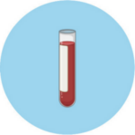
Blood test
Blood from the vein is drawn to identify antibodies
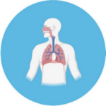
Lung function tests
Determine how well lungs function by measuring lung size and airflow in and out of the lungs
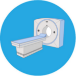
High resolution computed tomography (HRCT)
Creates detailed images of the inside of the chest and the characteristic PF pattern is shown as honeycombing
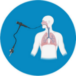
Bronchovascular lavage
A small flexible tube (bronchoscope) is used to sample cells from the lower part of the lungs (alveoli) for examination by a pathologist
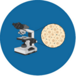
Thoracoscopic lung biopsy
A thin tube is placed to extract small samples of the lung from multiple lobes for testing in a laboratory
Content acknowledgements: © 2022 by the University of Hull and Authors. A Patient's Guide to Pulmonary Fibrosis Second Edition
